
But how easily could you convey such a sight without the latest digital camera? What if the most accurate method available was the fine point of engraver’s stylus?
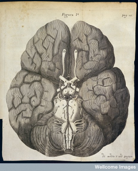
Written by an English physician named Thomas Willis this work was the first to give a complete account of how blood was supplied to the brain. The circulatory system that supports this most complex of organs is still known as the ‘Circle of Willis’ to this day. The Wellcome Library holds several editions of this seminal work complete with Wren’s original artwork.

Willis was also the first to use the word ‘neurology’ to describe the study of nerves. In his writings, he helped to focus on the solid tissues of the brain rather than the cavities and fluids emphasized in the more traditional humoral model of the body. His progressive ideas included attributing the cause of epilepsy to a physical condition rather than some form of ‘supernatural possession’, helping to pave the way for the research carried out by Robert Ludlow some 350 years later.
Images: Intracranial recording for epilepsy. Surface of human brain in situ 2011 (Wellcome Image no. N0036750)
The brain engraving from Cerebi anatome by Thomas Willis, published by J. Flesher for J. Martyn & J. Allestry, London 1664 (Wellcome Image no. L0018951)
Christopher Wren making his first demonstration of a method of introducing drugs into a vein, before Dr Willis, 1667. Oil painting by Ernest Board. (Wellcome Library no. 45901i)
No comments:
Post a Comment